Muscles Of The Chest And Abdomen Labeled - Chest and Arm Muscles Labeled Models - Biceps brachii ... - Check out this library of free labeling diagrams.
Muscles Of The Chest And Abdomen Labeled - Chest and Arm Muscles Labeled Models - Biceps brachii ... - Check out this library of free labeling diagrams.. Usually, the pain begins in the center of the chest, and it may radiate outward. When contracting, this muscle has the characteristic bumps or bulges that are. Labeling muscles (chest and abdomen). Fabian identifying the muscles and landmarks of the abdomen and chest. Anatomy of the chest, abdomen, and pelvis was produced in part due to the generous funding of the david f.
Ventral neck, chest and abdomen: The pectoantebrachialis has been separated from the underlying pectoralis major, and is being lifted in the image. Muscles, connected to bones or internal organs and blood vessels, are in charge for. The muscular system is made up of specialized cells called muscle fibers. Intercostal muscle strains are the most common cause of musculoskeletal chest pain, which people often refer to as a pulled muscle.
Female Muscle Diagram and Definitions | Jacki's Blog from aerobicdancing.com.au Free online quiz muscles of the chest and abdomen labeling. The abdomen (colloquially called the belly, tummy, midriff or stomach) is the part of the body between the thorax (chest) and pelvis, in humans and in other vertebrates. Muscles of the face, mouth, and pharynx. The pectoantebrachialis has been separated from the underlying pectoralis major, and is being lifted in the image. Much information can be gathered from simply watching the patient and looking at the abdomen. There are multiple functions of these chest muscles. There are three muscular layers of the abdominal wall, with a fourth layer in the middle anterior region. Extend your arms (and the band) fully in front of your chest, then.
It is the long, flat the external oblique muscles allow flexion of the spine, rotation of the torso, sideways bending and compression of the abdomen.
Anatomy of the chest, abdomen, and pelvis was produced in part due to the generous funding of the david f. The pectoantebrachialis has been separated from the underlying pectoralis major, and is being lifted in the image. This requires complete exposure of the region in question. The skeletal muscles of the abdomen form part of the abdominal wall, which holds and protects the gastrointestinal system. Labeling muscles (chest and abdomen). Primarily, there are three chest muscles involved in movement: The abdominal head of the pectoralis major muscle is one of three origins for the pectoralis major. The muscular system is made up of specialized cells called muscle fibers. Related online courses on physioplus. Their main function is contractibility. Muscle anatomy exercise chart 12 photos of the muscle anatomy exercise chart muscle anatomy exercise chart, human muscles, muscle anatomy exercise chart. Usually, the pain begins in the center of the chest, and it may radiate outward. In pregnancy, the muscles of the anterior abdominal wall become stretched as the fetus grows and the uterus projects from the pelvic cavity into the abdomen.
Intercostal muscle strains are the most common cause of musculoskeletal chest pain, which people often refer to as a pulled muscle. Innervation for muscles with chest wall attachments are labeled. Their main function is contractibility. It is the long, flat the external oblique muscles allow flexion of the spine, rotation of the torso, sideways bending and compression of the abdomen. Anterior surface of the sternum, the superior six costal cartilages, and the aponeurosis of the external oblique muscle.
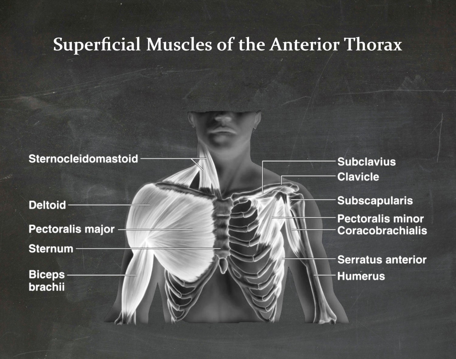
Muscles of the Chest - Muscles of the Anterior Thorax ... from img1.etsystatic.com Linea alba (white line of connective tissue at midline). Their main function is contractibility. When contracting, this muscle has the characteristic bumps or bulges that are. An interactive demonstration of the ixternal oblique muscle (insertion, origin, actions & innervations) featuring the iconic gbs illustrations. Muscles of the face, mouth, and pharynx. Abdominal muscles help you breathe out when you are breathing fast, such as during physical activity. One of the main smooth muscles inside the chest is the diaphragm. Here is the same image with the chest muscles labeled.
Topical anatomy of the abdomen.
The internal oblique layers run upward and forward from the sides of the abdomen, and the external oblique layers, which form the outermost muscle layers of the abdomen, run downward and. Extend your arms (and the band) fully in front of your chest, then. Muscle performance in neck pain online course: Here is the same image with the chest muscles labeled. Much information can be gathered from simply watching the patient and looking at the abdomen. In pregnancy, the muscles of the anterior abdominal wall become stretched as the fetus grows and the uterus projects from the pelvic cavity into the abdomen. Usually, the pain begins in the center of the chest, and it may radiate outward. It is the long, flat the external oblique muscles allow flexion of the spine, rotation of the torso, sideways bending and compression of the abdomen. Related online courses on physioplus. Common chest and abdominal injuries. The muscles of the anterior abdominal wall are located near the midline between the costal margin superiorly and the pubis inferiorly. One of the main smooth muscles inside the chest is the diaphragm. Muscular wall separating the chest and abdomen.
One of the main smooth muscles inside the chest is the diaphragm. Check out this library of free labeling diagrams. There are three muscular layers of the abdominal wall, with a fourth layer in the middle anterior region. Innervation for muscles with chest wall attachments are labeled. Muscles of the face, mouth, and pharynx.
Activity 2: Identifying Muscles of the Trunk Flashcards ... from www.easynotecards.com Check out this library of free labeling diagrams. Much information can be gathered from simply watching the patient and looking at the abdomen. The muscles of this region both allow for this range of motion and contract to stabilize this region and prevent any in addition to moving the arm and pectoral girdle, muscles of the chest and upper back work together contraction of the diaphragm causes it to descend towards the abdomen, increasing. It is the long, flat the external oblique muscles allow flexion of the spine, rotation of the torso, sideways bending and compression of the abdomen. Small muscles running between the ribs, known as the external intercostal muscles, lift the ribs during deep breathing to further expand the chest and lungs and provide even more air to the body. One of the main smooth muscles inside the chest is the diaphragm. Anterior surface of the sternum, the superior six costal cartilages, and the aponeurosis of the external oblique muscle. Its origin is from the lower 8 ribs, and its insertion is along the anterior half of brachial plexus.
Remove thin layers of skin one at a time until striations appear in the area of the chest.
Intercostal muscle strains are the most common cause of musculoskeletal chest pain, which people often refer to as a pulled muscle. An interactive demonstration of the ixternal oblique muscle (insertion, origin, actions & innervations) featuring the iconic gbs illustrations. The pectoralis major, the pectoralis minor, and the serratus anterior. The abdominal wall encloses the abdominal cavity, which holds the bulk of the gastrointestinal viscera. For some smaller muscle observations, larger. Innervation for muscles with chest wall attachments are labeled. The abdomen (colloquially called the belly, tummy, midriff or stomach) is the part of the body between the thorax (chest) and pelvis, in humans and in other vertebrates. Abdominal muscles help you breathe out when you are breathing fast, such as during physical activity. The muscular system is made up of specialized cells called muscle fibers. Muscular wall separating the chest and abdomen. The primary function is certainly to provide support to the skeletal system and to facilitate its movements. Check out this library of free labeling diagrams. Extend your arms (and the band) fully in front of your chest, then.
Common chest and abdominal injuries muscles of the chest abdomen. One of the main smooth muscles inside the chest is the diaphragm.


0 Komentar